Real-time OCT Berger´s space disection


images: http://www.ilustracionmedica.com

music: http://www.bensound.com

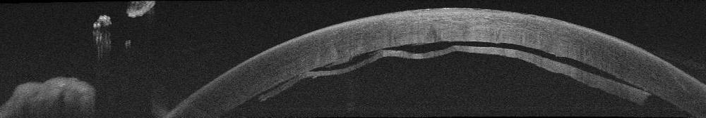
We report the case of a myopic patient with posterior subcapsular cataract secondary to several manipulations of the posterior segment . Usually it is not displayed by OCT intraoperative , but in this case is clearly seen the fibrous plaque in the posterior capsule , which breaks with the cystotome needle, separating the anterior hyaloid by injection of viscoelastic and placing the IOL in the capsular bag to occupy the physiological position of the lens.


Presentamos el caso de un paciente miope magno, con catarata subcapsular posterior secundaria a manipulaciones varias del segmento posterior. Habitualmente no se visualiza mediante OCT intraoperatorio, pero en este caso se observa con claridad la placa fibrosa en la cápsula posterior, que se rompe con un cistotomo, separando la hialoides anterior mediante inyección de viscoelástico y colocando la lente intraocular en el saco capsular, de manera que ocupe la posición fisiológica del cristalino.


Transfixing suture in DSAEK

We present a case of a patient with Fuchs’ corneal posterior dystrophy associated to a map-dot-fingerprint anterior dystrophy and a previously failed DMEK , which was treated with DSAEK . After 1 week , the graft began to peel off in an area that had previously suffered a traumatic abrasion of the predescemetic Dua´s layer . We decided to perform a transfixing suture asociated to rebubbleing to ensure the proper adhesion of the graft , since the latter was the third surgery in this patient . Tips and videos that can be seen explicitly showing the technique and the postoperative OCT. Presentamos un caso de una paciente con distrofia corneal posterior de Fuchs asociada a una distrofia anterior tipo mapa-punto-huella, y una DMEK fallida previa, en la que realizamos una DSAEK. Después de 1 semana, el injerto empezó a despegarse en una zona en la que previamente se había producido una abrasión traumática de la capa predescemética de Dua. Decidimos realizar un rebubbleing reforzado con puntos transfixiantes para asegurar la correcta adhesión del injerto, dado que ésta última era la tercera intervención quirúrgica a la que sometíamos a la paciente. Se muestran trucos y vídeos en los que se observa de forma explicita la técnica, así como el OCT postoperatorio.