SMCIS modified for hard cataracts. Tips

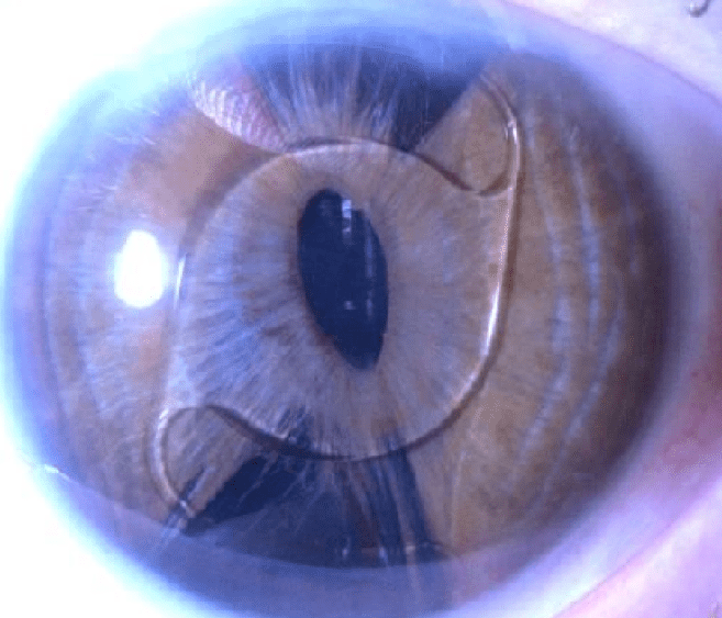
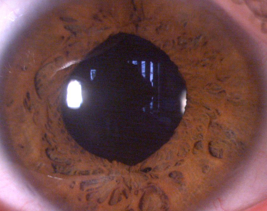
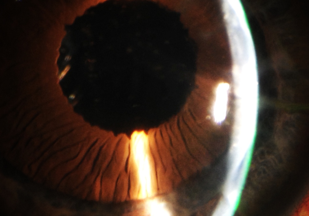
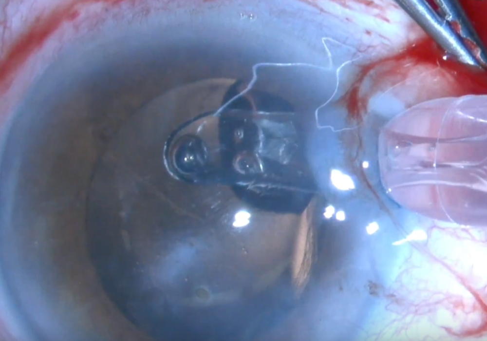
Small Incision Cataract Surgery (SMICS) is a very useful method to manage mature cataracts that could be a nightmare with Faco. This case is an only one eye with corneal leucoma an 4+ cataract. We add a limbal temporla incision to mantain the anteriro chamber tight and free of severe changes in the intraocular pressure but use the wide scleral tunnel to remove the cataract protecting the endothelium. We also perform a posterior capsulotomy during the surgery to ensure a good and quick visual rehabilitation and insert the IOL (Lucia from Zeiss) into the bag with the optic ocupying the capsulotomy and the haptics in the periphery. No vitrectomy is needed. The result is a safe and quick surgery with minimal endothelium damage (of course less that the one secondary to a faco in this eye) and a satisifed patient and surgeon.

Baikoff IOL removal + cataract surgery + pupilloplasty. How to do it without damaging the cornea and the iris

Baikoff angel-supported IOL was implantes during the 90´s to correct high myopia with good visual results but after 20 years we now must confront the fact that all these phakic intraocular lenses must be removed due to late complications like endothelial cell loss, iris-related complications, glaucoma and others like synechiae. Here is one example: a patient with ovalization of the pupil, halos, severe decrease of the endothelium cell population and inicial cataract.

This is one of the reasons why I like ICL from STAAR

iOCt in Lamellar Corneal Transplantation

After a long the working on it, finally there has been released an e-book that I am proud of: iOCT in lamellar corneal transplantation. This is the reason why the blog was on “pause” for some months. I leave you the link to see it.
Hope you enjoy it!!!
https://itunes.apple.com/es/book/oct-intraoperatoria-en-cirug%C3%ADa-lamelar-corneal/id1282060997?mt=11

http://kekulebooks.com/tienda/oct-intraoperatoria-en-cirugia-lamelar-corneal/

And soon in english!!!!

Real-time OCT Berger´s space disection


images: http://www.ilustracionmedica.com

music: http://www.bensound.com

We report the case of a myopic patient with posterior subcapsular cataract secondary to several manipulations of the posterior segment . Usually it is not displayed by OCT intraoperative , but in this case is clearly seen the fibrous plaque in the posterior capsule , which breaks with the cystotome needle, separating the anterior hyaloid by injection of viscoelastic and placing the IOL in the capsular bag to occupy the physiological position of the lens.


Presentamos el caso de un paciente miope magno, con catarata subcapsular posterior secundaria a manipulaciones varias del segmento posterior. Habitualmente no se visualiza mediante OCT intraoperatorio, pero en este caso se observa con claridad la placa fibrosa en la cápsula posterior, que se rompe con un cistotomo, separando la hialoides anterior mediante inyección de viscoelástico y colocando la lente intraocular en el saco capsular, de manera que ocupe la posición fisiológica del cristalino.