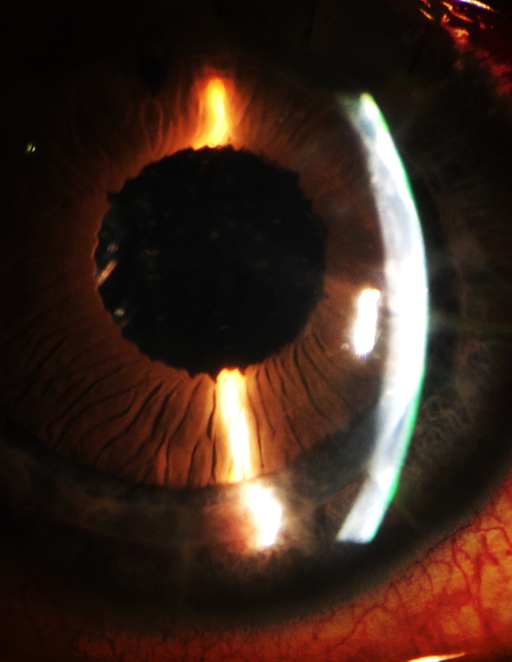
INTRACORNEAL RING REMOVAL 4 YEARS LATER


Challenging removal of a fibrotic and vascularised intracorneal segment. Four year after te implantation, the patient reports a white growing spot in the cornea. The Keraring is partially vascularised and covered by a fibrous tissue but not extruded. We tried several maneuvers to remove it, pulling from the hooked hole with an inverted simskey, pushing the distal area, or trying to dissect the primary incision… all of them useless. The only way to do it is looking at the previous records, making a new incision 2 mm from the end of the segment at the depthness set in the initial surgical plan and dissect with a blunt instrument over and under the segment, to refresh the tunnel and pull from one of the sides. No need of specific instruments, buy just in case, fill the tunnel with antibiotic and suture the incisions…

Baikoff IOL removal + cataract surgery + pupilloplasty. How to do it without damaging the cornea and the iris

Baikoff angel-supported IOL was implantes during the 90´s to correct high myopia with good visual results but after 20 years we now must confront the fact that all these phakic intraocular lenses must be removed due to late complications like endothelial cell loss, iris-related complications, glaucoma and others like synechiae. Here is one example: a patient with ovalization of the pupil, halos, severe decrease of the endothelium cell population and inicial cataract.

This is one of the reasons why I like ICL from STAAR

iOCt in Lamellar Corneal Transplantation

After a long the working on it, finally there has been released an e-book that I am proud of: iOCT in lamellar corneal transplantation. This is the reason why the blog was on “pause” for some months. I leave you the link to see it.
Hope you enjoy it!!!
https://itunes.apple.com/es/book/oct-intraoperatoria-en-cirug%C3%ADa-lamelar-corneal/id1282060997?mt=11

http://kekulebooks.com/tienda/oct-intraoperatoria-en-cirugia-lamelar-corneal/

And soon in english!!!!

Real-time OCT Berger´s space disection


images: http://www.ilustracionmedica.com

music: http://www.bensound.com

We report the case of a myopic patient with posterior subcapsular cataract secondary to several manipulations of the posterior segment . Usually it is not displayed by OCT intraoperative , but in this case is clearly seen the fibrous plaque in the posterior capsule , which breaks with the cystotome needle, separating the anterior hyaloid by injection of viscoelastic and placing the IOL in the capsular bag to occupy the physiological position of the lens.


Presentamos el caso de un paciente miope magno, con catarata subcapsular posterior secundaria a manipulaciones varias del segmento posterior. Habitualmente no se visualiza mediante OCT intraoperatorio, pero en este caso se observa con claridad la placa fibrosa en la cápsula posterior, que se rompe con un cistotomo, separando la hialoides anterior mediante inyección de viscoelástico y colocando la lente intraocular en el saco capsular, de manera que ocupe la posición fisiológica del cristalino.


Transfixing suture in DSAEK

We present a case of a patient with Fuchs’ corneal posterior dystrophy associated to a map-dot-fingerprint anterior dystrophy and a previously failed DMEK , which was treated with DSAEK . After 1 week , the graft began to peel off in an area that had previously suffered a traumatic abrasion of the predescemetic Dua´s layer . We decided to perform a transfixing suture asociated to rebubbleing to ensure the proper adhesion of the graft , since the latter was the third surgery in this patient . Tips and videos that can be seen explicitly showing the technique and the postoperative OCT. Presentamos un caso de una paciente con distrofia corneal posterior de Fuchs asociada a una distrofia anterior tipo mapa-punto-huella, y una DMEK fallida previa, en la que realizamos una DSAEK. Después de 1 semana, el injerto empezó a despegarse en una zona en la que previamente se había producido una abrasión traumática de la capa predescemética de Dua. Decidimos realizar un rebubbleing reforzado con puntos transfixiantes para asegurar la correcta adhesión del injerto, dado que ésta última era la tercera intervención quirúrgica a la que sometíamos a la paciente. Se muestran trucos y vídeos en los que se observa de forma explicita la técnica, así como el OCT postoperatorio.

Challenging cataract surgery

This is a 18 y-o patient who came to the emergency room refering a blunt trauma to her left eye. The patient had a corneal self-sealing wound, white cataract and rupture of the anterior capsule . Trypan blue staining is essential in these cases to locate the tear . Lens aspiration can be done with a Simcoe cannula, an angled cannula connected to a syringe or with the irrigation/aspiration tip . The idea is to keep the bag and anteriror hyalaoid to implant the lens in a physiological as possible position , maintaining the partitioning of the eye and looking the best postoperative visual acuity. Se trata de un paciente de 18 años de edad que acudió a urgencias por traumatismo contuso en ojo izquierdo. El paciente presentaba una herida corneal autosellada y catarata blanca por ruptura de la cápsula anterior. La tinción con azul tripan es esencial en estos casos para localizar el desgarro. La aspiración del cristalino se puede realizar con canula de Simcoe, con una cánula acodada conectada a una jeringa o bien con el tip de irrigacion aspiracion. La idea es mantener el saco y la hialaoides anterior íntegros para poder implantar la lente en una posición lo más fisiológica posible, manteniendo la compartimentación del ojo y buscando la mejor agudeza visual postoperatoria.

DSAEK after failed DMEK

This is a previously vitrectomized patient, with a failed DMEK and a severe cardiopathy. As DSAEK is more predictible in our hands, we perform it to treat the corneal edema and obtain a better visual acuity. Two recommendations: take off the DMEK graft under air and review the periphery looking for areas of angle clossure, as you will see in the superior anterior chamber of this patient with i-OCT . /Este es un paciente previamente vitrectomizado , con una DMEK fracasada y una cardiopatía grave. Puesto que en nuestras manoas es más predecible una DSAEK, la elegimos como tratamiento para el edema corneal y obtener una mejor agudeza visual. Dos recomendaciones : quitar el injerto DMEK bajo aire y revise la periferia en busca de áreas de cierre angular , como se verá en la cámara anterior superior de este paciente mediante i-OCT